Membrane Based Arrays (C-Series)
Membrane Based Antibody Arrays
Semi-Quantitative, Sandwich-Based, Membrane Antibody Arrays
The RayBio® Membrane-Based Antibody Arrays (C-Series) are tools for screening and comparing expression levels of many cytokines, growth factors, proteases, soluble receptors, and other proteins in a wide variety of sample types.
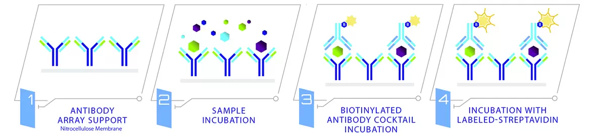
The C-Series Arrays utilize the sandwich immunoassay principle, wherein a panel of capture antibodies is printed on a nitrocellulose membrane solid support (usualy 2.5 cm x 3 cm). The array membranes are processed similarly to a Western blot (chemiluminescent readout). Signals are then visualized on x-ray film or a digital image, allowing densitometry data collection and calculation of fold-changes for each detected protein. The entire procedure can be completed in 1 day, and is simple enough to permit even the novice researcher to successfully collect data with very few pitfalls and little or no optimization.
Many biological processes such as apoptosis, inflammation, angiogenesis, immune response and migration often accompany changes of cytokine expression levels. Because of the extensive cross-talk between cytokines, a complete analysis of biological responses and functions must be obtained through multiplex assays. The C-Series Antibody Arrays allow a much broader view of protein activity than can be obtained with single-target ELISAs and Western blots. Moreover, antibody array screening improves the chances for discovering key factors, disease mechanisms or biomarkers related to cytokine signaling.
A variety of C-Series array kits are available, detecting anywhere from 10 to 274 proteins simultaneously.
Research Applications
- High-throughput profiling of cytokine expression
- Identifying potential molecular targets for drug development
- Identifying the molecular mechanisms of drug action
- Identifying crucial factors involved in disease processes
- Discovering biomarkers for disease management
- Discovering expression patterns for molecular classification of diseases
Features
- High Sensitivity: Can detect as low as 1 pg/ml for some proteins.
- High Reproducibility: Inter-array Coefficient of Variation (CV) of spot signal intensities as low as 5% when run under optimal conditions.
- High Specificity: A recombinant protein for each corresponding antibody spot is tested to ensure that only one particular spot is visible after hybridization.
- High Quality: Many antibody pairs are tested for each detectable protein target but only the pairs which show the strongest affinity and sensitivity are selected for the array.
- Wide Dynamic Range: A detectable range as high as 4 orders of magnitude (104) for some proteins.
- Robust Data Output: Each antibody is printed in duplicate providing multiple data points for each detected protein target.
- Positive & negative control spots: To orientate each membrane, monitor the detection steps, and normalize the results as well as to assess baseline and background responses.
- No dedicated equipment required: compatible with any chemiluminescent imaging system
- Easy to use: no training required
- Results can be obtained in 1 day
How It Works
Comparison to ELISA
- More Data, Less Sample: Antibody arrays provide high-content screening using about the same sample volume as for ELISA.
- Global View of Cytokine Expression: Antibody array screening improves the chances for discovering key factors, disease mechanisms or biomarkers related to cytokine signaling.
- Greater Sensitivity: As little as 4 pg/ml of MCP-1 can be detected using the C-Series array format. In contrast, our similar MCP-1 ELISA assay has a sensitivity of 40 pg/ml of MCP-1.
- Increased Range of Detection: ELISA assays typically detect a concentration range of 100- to 1000-fold, however, RayBiotech arrays can detect IL-2 at concentrations of 25 to 250,000 pg/ml, a range of 10,000-fold.
- Better Precision: As determined by densitometry, the inter-array Coefficient of Variation (CV) of spot signal intensities is 5-10%, comparing favorably with ELISA testing (CV = 10-15%).